 Coaching Centre
Discovering cells
How lenses revealed a hidden world: Robert Hooke and cork cells, Antonie van Leeuwenhoek's animalcules, the cell theory, unicellular and multicellular organisms, and why the electron microscope changed biology again.
In 1676 a Dutch cloth merchant sent a letter to the Royal Society in London. He said a single drop of rainwater, left to stand for a few days, was crowded with tiny creatures swimming about, so small that a million of them would not equal a grain of sand. The scientists thought he was exaggerating. It took them a year to build a microscope good enough to check. He was right.
Robert Hooke names the cell
Lenses for magnifying things had been around since the 1200s, mostly as reading glasses. Around 1590 spectacle makers in the Netherlands put two lenses in a tube and made the first compound microscope. By the 1660s scientists were using them to look at everything they could find.
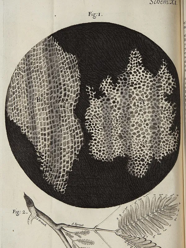
The English scientist Robert Hooke looked at fleas, feathers, mould and the point of a needle, and in 1665 he published his drawings in a book called Micrographia. One drawing showed a very thin slice of cork. It was full of little empty boxes, packed together like a honeycomb. Hooke called them cells, from the Latin cella, a small room, because they reminded him of the rooms monks lived in.
What Hooke saw were not living cells. Cork is bark, and its cells are dead: only their tough walls were left. He had no idea what cells did or that every living thing is made of them. But the name stuck.
Van Leeuwenhoek's animalcules
Antonie van Leeuwenhoek was the cloth merchant from the hook. He wanted to check the quality of threads, and taught himself to grind tiny, almost spherical lenses. His microscopes were nothing more than one bead of glass held in a brass plate, but his best lenses magnified about 270 times and gave a much sharper image than Hooke's.
From the 1670s he looked at pond water, rainwater, scrapings from his own teeth, blood and sperm. He was the first person to see single-celled organisms, which he called animalcules ("little animals"), and the first to see bacteria and red blood cells. He kept his lens-making method secret, so for a long time nobody could match him.
- Start with the pond water and your eye. Then step through each instrument. What can each one show that the one before could not?
- Watch the "smallest detail" number. Making things bigger only helps if the lens is sharp enough to show detail.
- Switch to the cork. Which instrument shows what Hooke drew in 1665?
The cell theory
For more than 150 years after Hooke, people knew cells existed but argued about what they were. Better lenses in the early 1800s settled it.
| Year | Scientist | What they showed |
|---|---|---|
| 1831 | Robert Brown | Saw a dark round structure in every plant cell he looked at and named it the nucleus. |
| 1838 | Matthias Schleiden | All plants are made of cells. |
| 1839 | Theodor Schwann | All animals are made of cells too, so cells are the basic unit of all living things. |
| 1855 | Rudolf Virchow | New cells only come from cells that already exist, by dividing. (Robert Remak had shown this first.) |
Together these ideas became the cell theory, one of the most important ideas in biology:
- All living things are made of one or more cells.
- The cell is the smallest unit that can carry out all the processes of life.
- All cells come from pre-existing cells.
The third point killed off an old belief called spontaneous generation: that maggots grow out of rotting meat or mice out of piles of grain. Living things only come from other living things.
One cell or many?
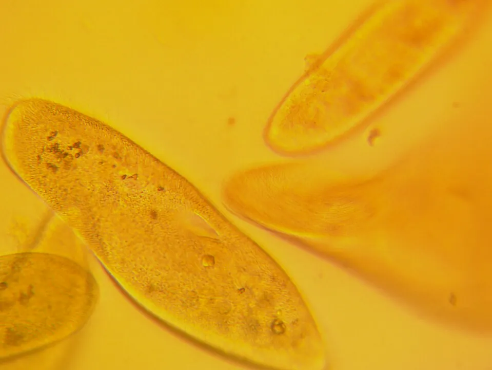
Some organisms are a single cell that does everything: feeds, gets rid of waste, responds and reproduces. These are unicellular organisms. Bacteria, the Paramecium and amoeba in pond water, and the yeast that makes bread rise are all unicellular.
Plants, animals and most fungi are multicellular: built from many cells that share out the work. In a multicellular organism, cells become specialised. A nerve cell can't digest food and a stomach cell can't carry a message to your brain, but together they keep the whole organism alive. You'll see how in topic 3G.
A second revolution: electron microscopes
Light microscopes have a limit. Light waves can't show details smaller than about 0.2 micrometres (a five-thousandth of a millimetre), however much you magnify. Viruses and the tiny structures inside cells stayed invisible.
In 1931 the German engineers Ernst Ruska and Max Knoll built the first electron microscope, which uses a beam of electrons instead of light. Modern electron microscopes magnify up to about 2 million times and show details a thousand times finer than a light microscope can.
- A transmission electron microscope (TEM) fires electrons through a very thin slice, showing the inside of a cell.
- A scanning electron microscope (SEM) bounces electrons off the surface, giving 3D-looking images of the outside of things.
There's a catch: specimens have to be in a vacuum, so they must be dead. Electron images are also black and white. The coloured pictures you see in books have been coloured on a computer.
Test yourself
Hooke chose the word "cell" because
Cella means a small room, like a monk's room in a monastery.
Van Leeuwenhoek could see bacteria and Hooke could not, mainly because van Leeuwenhoek's lenses
His tiny ground lenses gave clearer detail (better resolution), not just bigger images.
Which statement is not part of the cell theory?
Bacteria have no nucleus, and neither do your red blood cells.
A scientist wants a 3D-looking image of the surface of a pollen grain. The best instrument is a
An SEM bounces electrons off surfaces, giving detailed 3D-looking pictures.
Yeast is described as unicellular. This means it
"Uni" means one. Yeast is a single-celled fungus.
Put these discoveries in the order they happened.
One cell or many?
In 1665 Robert used a microscope to look at a slice of and called the little boxes cells. Van used single, carefully ground lenses to see living single-celled organisms. An organism made of just one cell is called . Light microscopes cannot show details smaller than about 0.2 .
Explain how improvements in technology led to the cell theory. Refer to at least two scientists.
Before microscopes, cells were too small to see, so nobody knew living things were made of them. Hooke's compound microscope showed the walls of cork cells in 1665, and van Leeuwenhoek's sharper lenses showed living single-celled organisms. In the 1800s even better lenses let Schleiden show that all plants are made of cells and Schwann show that animals are too. Their observations, together with Virchow's evidence that new cells come from dividing cells, became the cell theory.
Give one advantage and one disadvantage of an electron microscope compared with a light microscope.
An electron microscope can magnify up to about 2 million times and show much finer detail, such as viruses. However, the specimen must be dead because it is placed in a vacuum, so you cannot watch living cells.