 Coaching Centre
Cell size and specialised cells
Micrometres, the sizes of cells and the things inside them, using the field of view to estimate cell size, why cells are small, and how specialised cells such as nerve, red blood, sperm, muscle, root hair, guard and palisade cells are built for their jobs. Stem cells.
Your longest cells are nerve cells that run from the bottom of your spine to your big toe: about a metre long, but so thin you'd need a microscope to see one. Your smallest are red blood cells, 7 thousandths of a millimetre across, and you make about two and a half million new ones every second. Every cell's shape is a clue to the job it does.
Measuring tiny things: micrometres
Millimetres are far too big for cells. Scientists use the micrometre (µm), one thousandth of a millimetre. (The symbol µ is the Greek letter "mu".)
1 mm = 1000 µm so 1 µm = 0.001 mm
To change millimetres to micrometres, multiply by 1000. To change micrometres to millimetres, divide by 1000. Viruses are smaller still, and are measured in nanometres (nm): 1 µm = 1000 nm.
| Object | Approximate size |
|---|---|
| Flu virus | 0.1 µm (100 nm) |
| Staphylococcus bacterium | 1 µm |
| Mitochondrion | about 2 µm × 1 µm |
| E. coli bacterium | about 3 µm long |
| Chloroplast | about 5 µm × 2 µm |
| Red blood cell | 7 to 8 µm |
| Cheek cell | about 50 µm |
| Human egg cell | about 100 µm (just visible to the eye) |
| Onion skin cell | about 250 µm long |
Estimating size with the field of view
The circle of light you see down a microscope is the field of view. If you know how wide it is, you can estimate the size of anything in it.
To find it, put a clear plastic ruler on the stage at the lowest magnification (×40) and count the millimetre gaps across the circle. On most school microscopes it's about 4.5 mm, which is 4500 µm. At higher magnifications the field of view shrinks by the same factor as the magnification grows:
- ×40 → ×100 is 2.5 times more magnification, so the field of view is 4500 ÷ 2.5 = 1800 µm.
- ×40 → ×400 is 10 times more magnification, so the field of view is 4500 ÷ 10 = 450 µm.
At ×400 the field of view is 450 µm. About 9 cheek cells fit in a line across the middle of the circle.
Size of one cell ≈ field of view ÷ number of cells across = 450 ÷ 9 = 50 µm.
Check it makes sense: cheek cells are about 50 µm, so this is reasonable. If you got 50 000 or 0.05, you've mixed up millimetres and micrometres.
- Start with the ruler at ×40 and count the millimetre gaps. Then switch to ×100 and ×400. How does the field of view change?
- Pick onion cells. Choose a magnification where you can count them, count across the middle, then divide. Check your answer, then try "New sample".
- Try red blood cells. Which magnification do you need, and why?
Why are cells so small?
Everything a cell needs, such as oxygen and glucose, has to get in through its membrane, and wastes have to get out the same way. A small cell has a lot of membrane compared with the amount of cytoplasm inside it, and nothing inside is far from the edge. If a cell kept growing, its volume would increase faster than its surface, and substances couldn't move in and out fast enough to keep it alive. So instead of growing huge, cells divide. A blue whale isn't made of bigger cells than a mouse; it's made of more of them.
Specialised cells
All the cells in your body started from one fertilised egg, but as you developed they became specialised: each type has a shape and contents suited to one job.
| Cell | Job | How its structure helps |
|---|---|---|
| Nerve cell (neuron) | Carries electrical messages | Very long, thin fibre to carry messages over long distances; branched ends to connect with many other cells. |
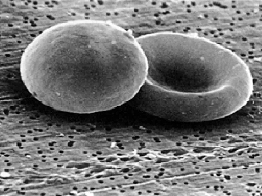
| Red blood cell | Carries oxygen | No nucleus, leaving more room for haemoglobin, which carries oxygen. Disc shape dented on both sides gives a big surface for oxygen to pass through. Small and flexible to squeeze through narrow capillaries. |
| Sperm cell | Reaches and fertilises an egg | Tail to swim. Many mitochondria to release energy for swimming. Head contains half the DNA needed for a new individual. |
| Muscle cell | Contracts to cause movement | Long and packed with protein fibres that slide together; many mitochondria. |
| Ciliated cell | Keeps airways clean | Tiny hairs (cilia) beat together to sweep mucus, with trapped dust and germs, up and out of the airways. |
| Root hair cell | Absorbs water and minerals | A long, thin extension gives a huge surface for absorbing water from soil. No chloroplasts, because there's no light underground. |
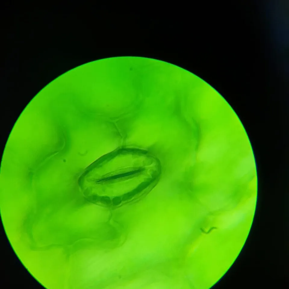
| Guard cells | Open and close pores in leaves | A pair of curved cells around each pore (stoma). They swell to open the pore and let gases in and out, and go limp to close it and save water. |
| Palisade cell | Photosynthesis | Tall, column-shaped cells near the top of the leaf, packed with chloroplasts to absorb as much light as possible. |
Stem cells
A stem cell is an unspecialised cell that can divide and turn into specialised cells. An early embryo is made almost entirely of stem cells that can become any cell type. Adults still have some, for example in bone marrow, where stem cells make about 100 billion new blood cells every day.
Doctors already use stem cells to treat some diseases. A bone marrow transplant gives a patient with leukaemia (a blood cancer) healthy stem cells that rebuild their blood. Blood from the umbilical cord, saved at birth, is another source. Researchers are now growing stem cells into heart muscle, nerve cells and even tiny model organs in the lab to test medicines and, one day, repair damaged tissue.
Test yourself
How many micrometres are in 2.5 mm?
2.5 × 1000 = 2500.
At ×40 the field of view is 4 mm. At ×400 it will be about
Ten times the magnification gives one tenth of the field of view.
The field of view is 1800 µm and 9 cells fit across it. Each cell is about
1800 ÷ 9 = 200 µm.
Which feature helps a root hair cell absorb water?
More surface touching the soil means more water can be absorbed.
Sperm cells contain lots of mitochondria because they
Mitochondria release the energy the tail needs.
Match each feature to its cell.
One micrometre is one of a millimetre. The circle you see down a microscope is called the field of . When the magnification increases, the field of view gets . A cell that is not yet specialised and can become many types of cell is a cell.
At ×100 the field of view of a microscope is 1.8 mm. A student counts 12 cells lined up across it. Estimate the length of one cell in micrometres. Show your working.
1.8 mm = 1.8 × 1000 = 1800 µm. Size of one cell ≈ 1800 ÷ 12 = 150 µm.
Explain how the structure of a red blood cell suits its function.
Red blood cells carry oxygen around the body. They have no nucleus, which leaves more space for haemoglobin, the substance that carries oxygen. Their disc shape, dented on both sides, gives a large surface area so oxygen can move in and out quickly, and they are small and flexible enough to squeeze through narrow capillaries.