 Coaching Centre
Mass spectrometry
How a mass spectrometer ionises, sorts and detects molecules; reading the molecular ion, base peak and fragments; and using isotope patterns for chlorine and bromine.
At an Olympic Games, a laboratory tests thousands of urine samples for banned substances, and each result has to stand up in court. The key instrument is a mass spectrometer. It can find nanograms of a steroid among thousands of natural compounds by breaking molecules into pieces and weighing each piece. Sport drug testing, airport explosive detectors and newborn-baby screening programs all rely on the same idea.
How a mass spectrometer works
Every mass spectrometer does four things, all in a high vacuum so the ions don't collide with air molecules:
- Ionisation. The vaporised sample is hit by a beam of high-energy electrons, which knock an electron off each molecule to form a positive ion, the molecular ion: $\text{M}(g) + e^- \rightarrow \text{M}^+(g) + 2e^-$. The molecular ion has so much spare energy that many of them break apart (fragment) into a smaller positive ion and a neutral piece.
- Acceleration. An electric field accelerates the positive ions to high speed.
- Separation. The ions are sorted by their mass-to-charge ratio, $m/z$. In the classic design, a magnetic field bends the ions: light ions are deflected more than heavy ones. Since almost all ions have a charge of +1, $m/z$ is simply the mass of the ion.
- Detection. A detector counts the ions at each $m/z$, and a computer plots the mass spectrum.
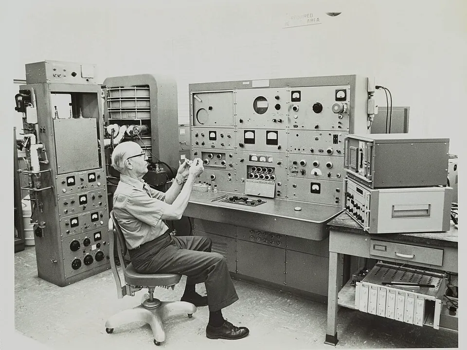
Textbook diagrams usually show the magnetic-sector design, and it is the one to describe in an exam. Most modern instruments separate ions instead by their time of flight along a tube (lighter ions arrive first) or with a quadrupole of oscillating electric fields. Many are coupled to a gas or liquid chromatograph, which separates a mixture first so each compound reaches the mass spectrometer on its own.
Reading a mass spectrum
- The molecular ion peak, M⁺, is the peak at the highest $m/z$ (ignoring tiny isotope peaks). Its $m/z$ equals the relative molecular mass. For ethanol it is 46.
- The base peak is the tallest peak, set to 100%. It comes from the most abundant ion, usually the most stable fragment. For ethanol it is CH₂OH⁺ at 31.
- The fragment peaks show which pieces the molecule broke into. The difference between M⁺ and a fragment tells you the neutral piece that was lost. For ethanol, $46 - 31 = 15$, the mass of a CH₃ group.
Only the charged piece of each fragmentation is detected; the neutral piece is invisible. A molecule can break in several places, so you often see both CH₃⁺ (15) and the ion left after losing CH₃ (M − 15).
| Mass | As an ion: a peak at this m/z | As a neutral loss: a peak at M − this mass |
|---|---|---|
| 15 | CH₃⁺ | lost CH₃ |
| 17 | – | lost OH |
| 18 | – | lost H₂O (common for alcohols) |
| 29 | C₂H₅⁺ or CHO⁺ | lost C₂H₅ or CHO |
| 31 | CH₂OH⁺ (primary alcohols) | lost OCH₃ or CH₂OH |
| 43 | CH₃CO⁺ or C₃H₇⁺ | lost C₃H₇ |
| 45 | COOH⁺ | lost COOH |
An unknown with formula C₃H₆O has a molecular ion at m/z 58, a base peak at 43 and a small peak at 15. There is no significant peak at 29. Is it propanone, CH₃COCH₃, or propanal, CH₃CH₂CHO?
Isotope peaks
Because the spectrometer weighs individual ions, molecules made with different isotopes appear at different $m/z$:
- Chlorine is about 75% ³⁵Cl and 25% ³⁷Cl, so a compound with one Cl atom shows peaks at M and M + 2 in a 3 : 1 ratio.
- Bromine is about 51% ⁷⁹Br and 49% ⁸¹Br, so one Br atom gives M and M + 2 peaks of almost equal height.
- Carbon-13 (1.1% of carbon) gives a small M + 1 peak. Its height grows with the number of carbon atoms.
An M + 2 pattern is therefore a quick way to spot a halogen that no other technique on this page shows so clearly.
- For ethanol, step to m/z 31 and confirm that 46 − 31 = 15 (loss of CH₃). Then compare propan-2-ol: which peak is its base peak, and what was lost?
- Compare propanone and butanone. Both have a base peak at 43. Use the molecular ions (58 and 72) to work out what each lost to form CH₃CO⁺.
- Open chloroethane. Find the two molecular ion peaks (64 and 66) and check that their heights are in about a 3 : 1 ratio.
- Try three mystery spectra. Use only M⁺ and the base peak to make your first guess, then check the other peaks.
Test yourself
In a mass spectrometer, ions are separated according to their
Deflection (or flight time) depends on m/z; most ions have z = 1.
The mass spectrum of a compound has its molecular ion at m/z 74 and a strong peak at m/z 45. The neutral fragment lost to form the m/z 45 ion has a mass of
74 − 45 = 29, which matches C₂H₅. Propanoic acid losing C₂H₅ leaves COOH⁺.
A compound shows two molecular ion peaks at m/z 108 and 110 of almost equal height. It most likely contains
⁷⁹Br and ⁸¹Br are about equally abundant. (Bromoethane, C₂H₅Br, has M = 108 and 110.)
The base peak in a mass spectrum is the peak
The tallest peak is set to 100% and comes from the most common ion.
Name each stage of the mass spectrometer.
Put the stages of mass spectrometry in order.
The peak at the highest m/z is the peak and gives the relative molecular mass. The tallest peak is called the peak. Only ions are detected. A compound containing one chlorine atom shows M and M + 2 peaks in a ratio of about .
The mass spectrum of an alcohol with formula C₃H₈O has peaks at m/z 60 (small), 45 (100%) and 43. Explain how the spectrum shows whether the compound is propan-1-ol or propan-2-ol.
The molecular ion at m/z 60 matches C₃H₈O, so it fits both isomers. The base peak at 45 is 60 − 15, so the most common fragmentation is loss of a CH₃ group. In propan-2-ol, CH₃CH(OH)CH₃, the carbon holding the OH has two CH₃ groups attached; losing one leaves CH₃CHOH⁺, m/z 45, which is stabilised by the oxygen. Propan-1-ol, CH₃CH₂CH₂OH, would mainly break next to its CH₂OH to lose C₂H₅ (29), giving CH₂OH⁺ at m/z 31 as the base peak. The base peak at 45 (and not 31) therefore identifies propan-2-ol.