 Coaching Centre
Infrared spectroscopy
Why covalent bonds absorb infrared radiation, how to read an IR spectrum using wavenumber ranges from the data sheet, and how to tell alcohols, carboxylic acids, carbonyl compounds and alkenes apart.
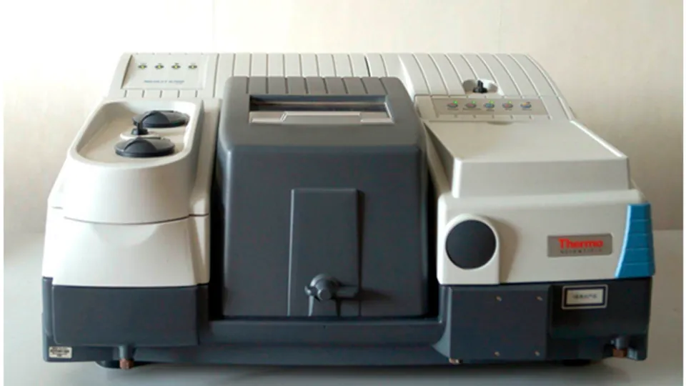
A roadside breath test for alcohol uses a fuel cell, but the evidential breath-analysis instrument at a police station often uses infrared light. Ethanol's C–H and C–O bonds absorb infrared at particular frequencies, so the amount of IR that a breath sample absorbs shows how much ethanol it contains. The same idea lets a chemist see which bonds are in an unknown molecule.
Why bonds absorb infrared
A covalent bond behaves like a spring joining two masses. It can stretch and bend, and each kind of vibration has a natural frequency. When infrared radiation of exactly that frequency passes through, the bond absorbs it and vibrates more strongly. The frequency depends on:
- bond strength: stronger bonds vibrate faster, so C≡C absorbs at a higher frequency than C=C, which is higher than C–C;
- the masses of the atoms: bonds to light hydrogen atoms (O–H, N–H, C–H) vibrate fastest and absorb at the highest frequencies.
A vibration only absorbs IR if it changes the molecule's dipole. That is why polar bonds such as C=O and O–H give strong absorptions, and why N₂ and O₂ don't absorb IR at all. It is also why CO₂, H₂O and CH₄ are greenhouse gases: their vibrations absorb the infrared radiation emitted by Earth's surface.
Reading an IR spectrum
An IR spectrum plots transmittance (the percentage of light that gets through) against wavenumber, the number of waves per centimetre, in cm⁻¹. Wavenumber is proportional to frequency and energy. By convention the axis runs from 4000 cm⁻¹ on the left down to about 500 cm⁻¹, and absorptions appear as dips pointing downwards.
- Above 1500 cm⁻¹ are the absorptions of particular bonds. These identify functional groups.
- Below about 1500 cm⁻¹ is the fingerprint region: many overlapping absorptions from the whole molecule. You don't assign these, but the pattern is unique, so it can be matched to a database spectrum to confirm an identity.
| Bond | Wavenumber (cm⁻¹), from the HSC data sheet | What it looks like |
|---|---|---|
| O–H (alcohols) | 3230–3550 | strong, broad (hydrogen bonding) |
| N–H (amines) | 3300–3500 | medium; two sharp peaks for –NH₂ |
| C–H | 2850–3300 | in almost every organic compound |
| O–H (acids) | 2500–3000 | very broad, overlapping the C–H peaks |
| C≡N | 2220–2260 | sharp |
| C=O | 1680–1750 | strong, sharp: usually the most obvious peak |
| C=C | 1620–1680 | medium to weak |
| C–O | 1000–1300 | strong, in the fingerprint region |
| C–C | 750–1100 | weak, rarely useful |
Telling functional groups apart
A good routine is to ask two questions:
- Is there a strong, sharp C=O peak near 1700 cm⁻¹? If yes, the compound is an aldehyde, ketone, carboxylic acid, ester or amide.
- Is there a broad O–H band? A broad band centred around 3300 cm⁻¹ means an alcohol. A very broad band from 2500 to 3300 cm⁻¹ that swamps the C–H peaks means a carboxylic acid, especially if C=O is also present.
So an alcohol has O–H but no C=O; a ketone or ester has C=O but no O–H; a carboxylic acid has both. An aldehyde can also show two weak C–H peaks near 2720 and 2820 cm⁻¹. IR shows which bonds are present but rarely how they are arranged, so propan-1-ol and propan-2-ol look almost identical above 1500 cm⁻¹.
- Tap the broad dip in ethanol's spectrum. It reads at about 3350 cm⁻¹. Which data sheet range includes it?
- Compare ethanol, ethanoic acid and propanone. Which has O–H only, which has C=O only, and which has both?
- Find the C=C absorption in hex-1-ene (about 1640 cm⁻¹). Compare its depth with propanone's C=O. Why is the C=O so much stronger?
- Open ethylamine and find its two N–H peaks. Then try three mystery spectra, using the data-sheet overlay only to check your answer.
Test yourself
An IR spectrum has a strong, sharp absorption at 1715 cm⁻¹ and no broad absorption above 2500 cm⁻¹. The compound could be
C=O is present but O–H is not, which fits a ketone.
Why does nitrogen gas, N₂, not absorb infrared radiation?
N₂ is non-polar whether stretched or not, so it can't interact with IR.
The region of an IR spectrum below about 1500 cm⁻¹ is mainly used to
The fingerprint region is unique, so it is matched against databases.
Which pair of absorptions best identifies a carboxylic acid?
An acid shows both the very broad acid O–H and a C=O.
Name each labelled absorption or region.
Sort the compounds by the key features of their IR spectra.
IR spectra are plotted against , in cm⁻¹. The region below 1500 cm⁻¹ is called the region. Hydrogen bonding makes the O–H absorption . A bond only absorbs IR if its vibration changes the molecule's .
A compound X has the formula C₃H₆O₂. Its IR spectrum has a very broad absorption from 2500 to 3300 cm⁻¹ and a strong absorption at 1710 cm⁻¹. A second compound Y, with the same formula, has a strong absorption at 1740 cm⁻¹ and nothing above 3000 cm⁻¹. Identify the functional group in each and suggest a structure for each.
X: the very broad absorption from 2500 to 3300 cm⁻¹ lies in the O–H (acid) range of 2500–3000 cm⁻¹, and the strong absorption at 1710 cm⁻¹ is in the C=O range of 1680–1750 cm⁻¹. Having both means a carboxylic acid; with C₃H₆O₂ it is propanoic acid, CH₃CH₂COOH.
Y: the strong 1740 cm⁻¹ absorption shows C=O, but there is no O–H band, so it is not an acid or alcohol. With two oxygen atoms and one C=O, it is an ester: methyl ethanoate, CH₃COOCH₃, or ethyl methanoate, HCOOCH₂CH₃. IR alone can't tell these two apart; NMR could.